Microscopy Quiz
Introduction to Microscopy: Unveiling the Hidden World
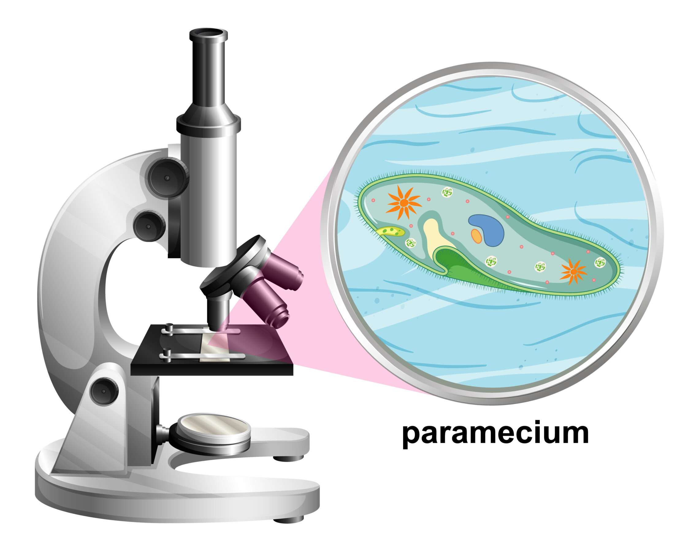
The quintessential tool for biologists, the microscope has long been a cornerstone in the realm of scientific exploration. The inaugural compound microscope, a groundbreaking invention, emerged in 1590, credited to the ingenuity of Zacharias Jenssen in Holland.

Microscopy Fundamentals: Magnification and Resolution
A microscope, the instrument of choice for scrutinizing minuscule entities, opens the gateway to the world of microscopy, where magnification and resolution take center stage. The augmentation of an object’s apparent size falls under the umbrella term “magnification,” while the resolving power, or resolution, denotes an optical instrument’s ability to differentiate two distinct points. This critical measure is defined as the minimum distance required for two points to be perceived as separate entities. The augmentation of resolution is made possible through the strategic use of lenses, a stark contrast to the innate human eye, which boasts a resolution limit of 0.1 millimeter.
Light Microscope vs. Electron Microscope: Illuminating the Differences

Diving into the dichotomy of microscopy, we encounter two prominent players: the light microscope and the electron microscope. The former harnesses visible light to illuminate objects, boasting eyepiece and objective lenses for enhanced viewing. With a magnification potential of 1500X and a resolution capacity of 0.2 micrometers, the light microscope is a workhorse in biological exploration.
On the other end of the spectrum, the electron microscope ushers in a new era by employing a fine electron beam transmitted to specimens in a vacuum. Operating with electromagnets as lenses, this advanced microscope generates images on a screen. Pushing the boundaries, the electron microscope achieves staggering magnifications of up to 1,000,000X and a remarkable resolution of 0.2 nanometers.
Electron Microscopy: A Closer Look at TEM and SEM
- Delving into the world of electron microscopy and its advanced capabilities.
- Introducing Transmission Electron Microscope (TEM) and its application in studying internal cell structures, involving thin specimen sections.
- Exploring Scanning Electron Microscope (SEM) and its focus on surface analysis, revealing three-dimensional intricacies.

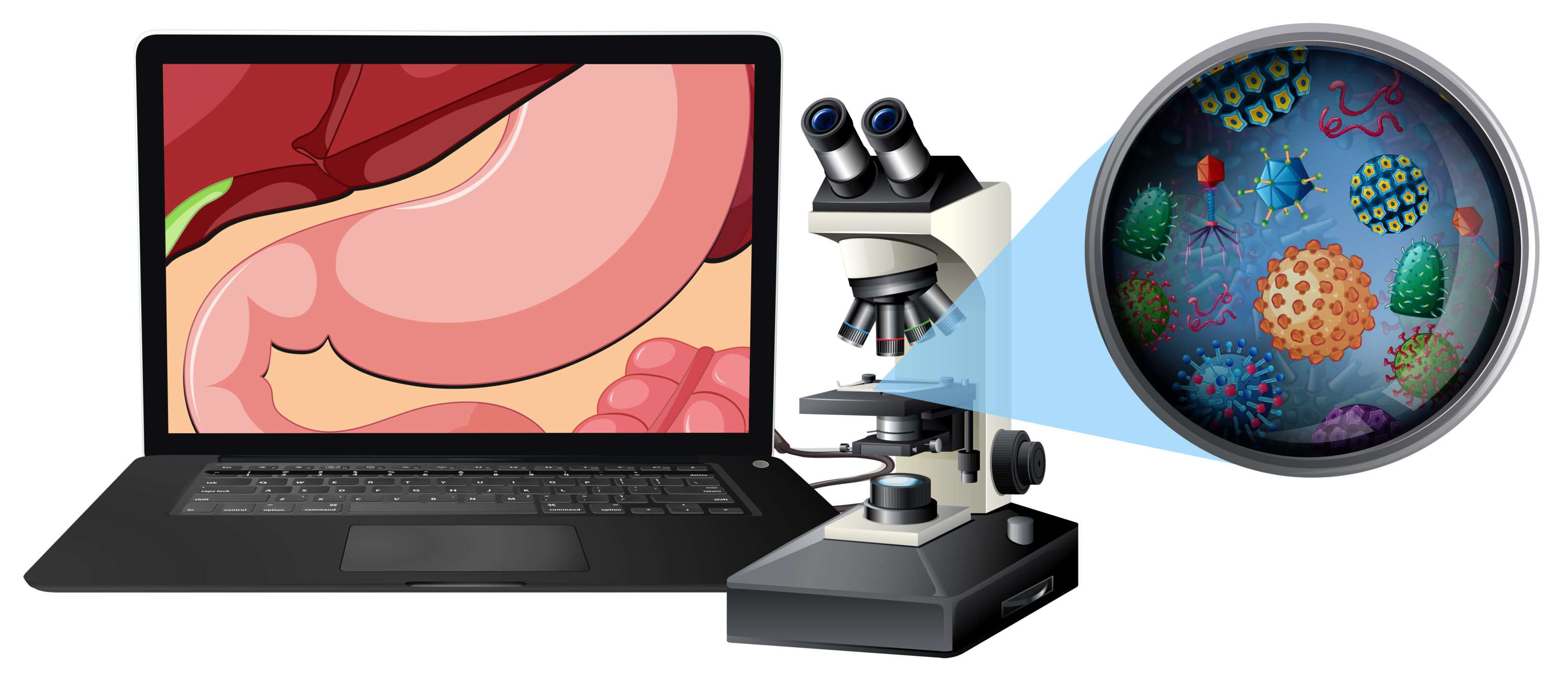
Go Check out and Learn: MCQs on Stomach Anatomy


