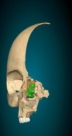
Welcome to a fascinating exploration of the cranial bones that make up the human skull. In this post, we’ll take you on a journey through these important bones, each thoughtfully labeled for clarity.
The skull is divided into two main sections: the cranial bones and the facial bones. The cranial bones encase and shield the brain, forming a protective layer around its delicate tissues. On the other hand, the facial bones form the framework of the face, supporting the eyes, nose, mouth, and other sensory features.
Here we will explore the cranial bones so let’s start to explore them.

Frontal Bone: (01)
The frontal bone is a single bone located at the front of the skull.

Location:
It forms the forehead region and extends posteriorly to contribute to the upper part of the eye sockets, known as the orbits.
Nasal Cavity:
The frontal bone also plays a role in the formation of the roof of the nasal cavity.
Protection:
The frontal bone provides protection to the underlying frontal lobes of the brain, one of the major regions responsible for cognitive functions.
Suture:
The frontal bone articulates with the parietal bones at the coronal suture, forming a junction between the frontal and parietal regions of the skull.
External Features:
The external surface of the frontal bone is generally smooth and convex, contributing to the characteristic shape of the forehead.
Sinuses:
The frontal bone contains frontal sinuses, and air-filled cavities that are part of the paranasal sinuses system.
Development:
During early development, the frontal bone consists of separate right and left halves that fuse together along the midline to form a single bone.
Blood Supply:
The frontal bone receives blood supply from branches of the ophthalmic and superficial temporal arteries.
Cranial Nerves:
While not directly housing any cranial nerves, the frontal bone provides a protective enclosure for several important nerves that pass through the skull, such as the olfactory nerves responsible for the sense of smell.
Parietal Bones: (02)

Parietal Bones: The skull consists of two parietal bones, one on each side, which are paired and symmetrical.
Location:
The parietal bones are positioned on the sides and upper part of the skull, forming the majority of the cranial vault.
Suture:
The parietal bones meet at the midline of the skull and are connected by the sagittal suture. This suture runs from the anterior (front) to the posterior (back) part of the skull.
Function:
The parietal bones provide protection and support to the sides of the brain, forming a sturdy enclosure for this vital organ.
Articulations:
The parietal bones articulate with other bones of the skull, including the frontal bone at the front, the occipital bone at the back, and the temporal bones at the sides.
External Features:
The external surface of the parietal bones is typically smooth and convex, contributing to the rounded shape of the skull.
Blood Supply:
The parietal bones receive blood supply from branches of the middle meningeal artery, which is an important vascular structure in the head.
Cranial Nerves:
The parietal bones do not directly house any cranial nerves but provide protection and support for these nerves as they pass through the cranial cavity.
Development:
The parietal bones initially develop from two separate bones during early development and gradually fuse together along the sagittal suture as a person grows.
Individual Variations:
The parietal bones can exhibit individual variations in shape, size, and surface features, although they generally follow the characteristic pattern of the parietal bones.
Temporal Bones: (02)
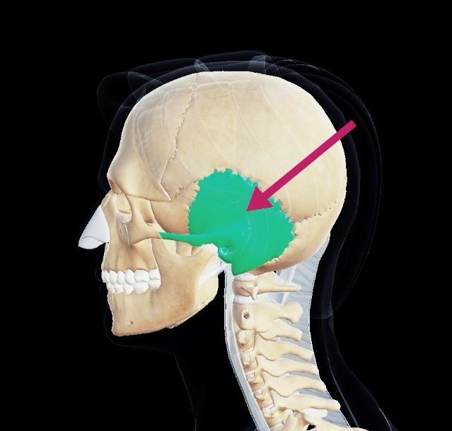
The skull consists of two temporal bones, one on each side, located on the sides and base of the skull.
Location:
The temporal bones are situated above the ear and extend to the lower sides of the skull.
Sutures:
The temporal bones articulate with other bones of the skull, including the parietal bones (via the squamosal sutures) and the sphenoid bone (via the sphenosquamosal sutures).
External Features:
The temporal bones have several notable external features, including the zygomatic process, which contributes to the formation of the cheekbone, and the external auditory meatus, which is the opening of the ear canal.
Internal Features:
Within the temporal bones, there are important structures such as the middle and inner ear cavities, which house components of the auditory system, including the ossicles (small bones involved in hearing) and the cochlea (involved in sound transmission).
Mandibular Fossa and Styloid Process:
The temporal bones feature the mandibular fossa, a depression that articulates with the condyle of the mandible (lower jawbone), forming the temporomandibular joint (TMJ). Additionally, the temporal bones contain the elongated styloid process, which serves as an attachment point for various muscles and ligaments.
Mastoid Process:
The mastoid process is a prominent bony projection located behind the ear. It provides attachment for certain neck muscles and is often palpable.
Petrous Portion:
The temporal bones encompass the petrous portion, which houses the delicate structures of the inner ear, including the cochlea, semicircular canals, and vestibule.
Blood Supply:
Blood supply to the temporal bones is provided by branches of the external carotid artery, including the middle meningeal artery.
Cranial Nerves:
The temporal bones contain important foramina (openings) through which cranial nerves pass, including the facial nerve (VII), the vestibulocochlear nerve (VIII), and the glossopharyngeal nerve (IX).
Occipital Bone (01):
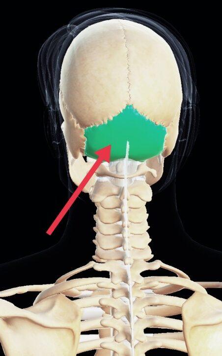
The occipital bone is a single bone located at the back of the skull, forming the posterior (back) part of the cranial vault.
Location:
It is positioned in the lower part of the skull, posterior to the parietal bones and temporal bones.
Foramen Magnum:
One of the most prominent features of the occipital bone is the foramen magnum, a large opening through which the spinal cord passes. This opening connects the cranial cavity with the spinal canal.
Articulations:
The occipital bone articulates with other bones of the skull, including the parietal bones at the sides, the temporal bones at the sides and base, and the sphenoid bone in front.
External Features:
The external surface of the occipital bone is marked by various bony prominences, such as the external occipital protuberance and superior and inferior nuchal lines, which serve as attachment sites for muscles and ligaments.
Internal Features:
Internally, the occipital bone contains important structures, including the occipital condyles. These rounded surfaces articulate with the first cervical vertebra (atlas), allowing for nodding movements of the head.
Function:
The occipital bone plays a crucial role in protecting the back of the brain and supporting the weight of the skull.
Development:
During fetal development, the occipital bone forms from several separate bones that gradually fuse together as the individual grows.
Blood Supply:
The occipital bone receives blood supply from branches of the occipital artery, which is a branch of the external carotid artery.
Cranial Nerves: While the occipital bone does not directly house any cranial nerves, it provides passage for cranial nerves that emerge from the brainstem and passes through the foramen magnum.
Sphenoid Bone: (01)

The sphenoid bone is a complex bone located at the base of the skull, behind the eye sockets.
Butterfly Shape:
The sphenoid bone has a unique butterfly-like shape, with a central body and extending wings.
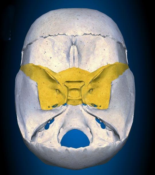
Keystone Bone:
The sphenoid bone is often referred to as the “keystone” bone because it articulates with many other bones of the skull, providing stability and structural support.
Cranial Floor:
The sphenoid bone forms a significant portion of the cranial floor, contributing to the structural integrity and shape of the skull base.
Eye Socket Formation:
The sphenoid bone plays a crucial role in forming the posterior part of the eye sockets (orbits), housing and protecting the delicate structures of the eyes.
Sphenoidal Sinuses:
Within the body of the sphenoid bone, there are air-filled spaces called sphenoidal sinuses. These sinuses help reduce the overall weight of the skull.
Optic Canal and Superior Orbital Fissure:
The sphenoid bone contains openings such as the optic canal and superior orbital fissure, which allow the passage of important structures like the optic nerve and blood vessels.
Sella Turcica:
Located on the superior surface of the sphenoid bone, the sella turcica is a bony saddle-shaped structure that houses the pituitary gland.
Foramen Rotundum, Foramen Ovale, and Foramen Spinosum:
The sphenoid bone has various foramina (openings) that allow the passage of nerves, blood vessels, and connective tissues within the skull.
Cranial Nerve Connections:
The sphenoid bone serves as a connection point for several cranial nerves, including the optic nerve (cranial nerve II) and the oculomotor nerve (cranial nerve III).
Ethmoid Bone: (01)
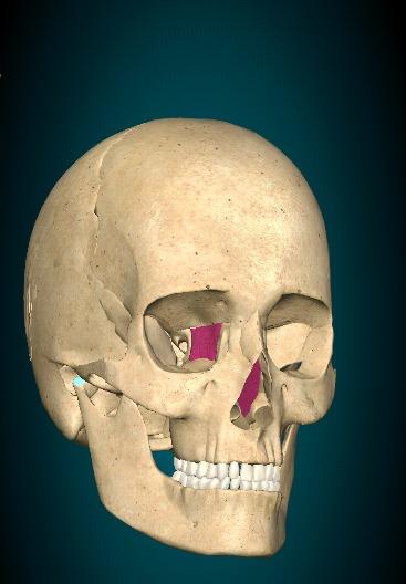
The ethmoid bone is a delicate and intricate bone located within the skull’s central area, contributing to the structure of both the cranium and the face.
Location and Divisions:
The ethmoid bone is situated between the eyes, forming a part of the nasal cavity’s walls and the nasal septum. It’s divided into two main parts: the vertical cribriform plate and the horizontal perpendicular plate.
Nasal Cavity and Nasal Septum:
The ethmoid bone’s cribriform plate forms the roof of the nasal cavity and contains small holes (olfactory foramina) through which the olfactory nerves pass, allowing for the sense of smell. The perpendicular plate contributes to the nasal septum, dividing the nasal cavity into left and right sides.
Cranial Base:
The ethmoid bone contributes to the anterior cranial fossa, which is one of the three depressions on the base of the cranial cavity that supports the frontal lobes of the brain.
Orbital Structures:
The ethmoid bone also plays a role in forming the medial walls of the eye sockets (orbits). It contributes to the ethmoidal labyrinth, which is a series of delicate, thin-walled air cells that help reduce the skull’s weight while still maintaining its strength.
Olfactory Nerves:
The ethmoid bone houses the olfactory bulb, where the olfactory nerves (cranial nerve I) arise. These nerves are essential for the sense of smell and pass through the cribriform plate to connect with the olfactory centers in the brain.
Air Circulation and Humidification:
The ethmoidal labyrinth’s air cells help to humidify and regulate the temperature of the inhaled air as it passes through the nasal passages. This contributes to the overall respiratory function.
Development:
During embryonic development, the ethmoid bone forms from multiple ossification centers that gradually fuse together. This intricate process results in the unique and complex structure of the bone.
Blood Supply:
The ethmoid bone receives blood supply from branches of the ophthalmic artery, which is a branch of the internal carotid artery.
Sinuses:
The ethmoid bone contains ethmoidal sinuses, which are a collection of air-filled cavities that are part of the paranasal sinuses system. These sinuses play a role in resonating the voice and reducing the overall weight of the skull.