The structure of heart is one of the most fascinating aspects of human anatomy. The heart is a muscular organ that functions as the central pump of the circulatory system, ensuring continuous blood flow throughout the body. Understanding the human heart structure is essential for students, medical aspirants, and anyone interested in how life is sustained.
Quiz
Available options: 1 to 20
In this article, we will explore the anatomy of the heart, its chambers, valves, layers, and blood circulation, in detail.
What is the Structure of Heart?
The human heart is a hollow, muscular organ about the size of a clenched fist. It is located slightly left of the midline in the chest, protected by the ribcage. The structure of heart is designed to pump oxygen-rich blood to the body and return oxygen-poor blood to the lungs.
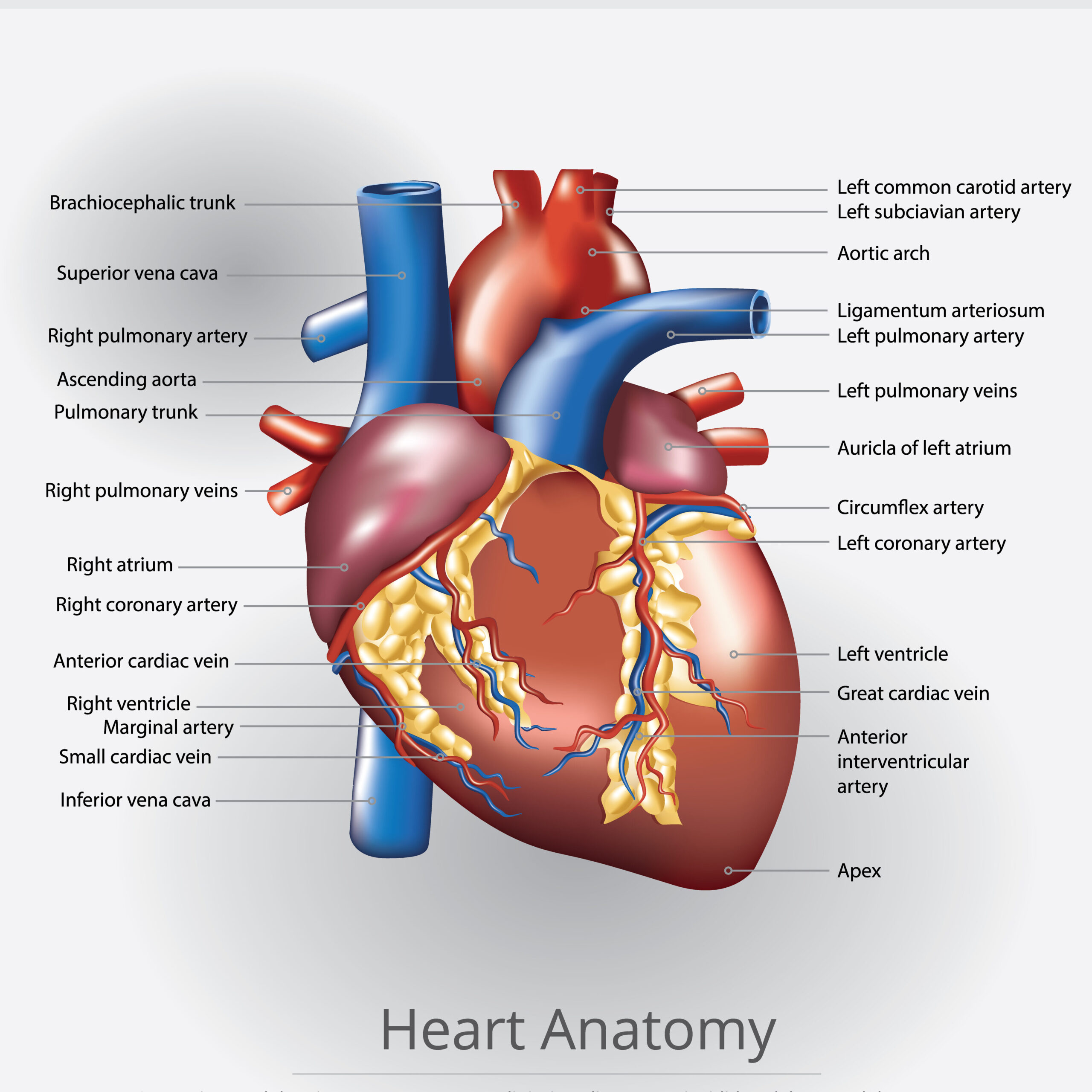
External Structure of Heart
From the outside, the structure of heart appears as a cone-shaped organ with major blood vessels attached. These include:
- Aorta – the largest artery carrying oxygenated blood.
- Pulmonary arteries – transport deoxygenated blood to the lungs.
- Pulmonary veins – return oxygenated blood to the heart.
- Vena cava (superior and inferior) – bring deoxygenated blood from the body back to the heart.
The heart is enclosed in a pericardium, a double-layered sac that protects it and reduces friction during beating.
Internal Structure of Heart
The internal structure of the heart is divided into four chambers:
1. Right Atrium
- Receives deoxygenated blood from the superior and inferior vena cava.
- Pumps blood into the right ventricle through the tricuspid valve.
2. Right Ventricle
- Receives blood from the right atrium.
- Pumps deoxygenated blood into the pulmonary arteries through the pulmonary valve.
3. Left Atrium
- Receives oxygen-rich blood from the pulmonary veins.
- Pushes blood into the left ventricle through the bicuspid (mitral) valve.
4. Left Ventricle
- The strongest chamber of the heart.
- Pumps oxygenated blood into the aorta through the aortic valve.
Valves of the Heart
The structure of heart includes four valves that regulate the flow of blood and prevent backflow:
- Tricuspid valve – between right atrium and right ventricle.
- Pulmonary valve – between right ventricle and pulmonary artery.
- Mitral valve – between left atrium and left ventricle.
- Aortic valve – between left ventricle and aorta.
These valves ensure unidirectional blood flow.
Layers of the Heart Wall
The wall of the heart structure has three layers:
- Epicardium – outer protective layer.
- Myocardium – thick, muscular middle layer responsible for contractions.
- Endocardium – smooth inner layer lining the chambers.
Blood Circulation in the Heart
The heart structure is designed to maintain a double circulation:
- Pulmonary circulation: Right side of the heart sends deoxygenated blood to the lungs.
- Systemic circulation: Left side of the heart pumps oxygenated blood to the rest of the body.
This ensures continuous supply of oxygen and removal of carbon dioxide.
Functions of the Structure of Heart
- Pumping blood throughout the body.
- Supplying oxygen and nutrients to tissues.
- Removing carbon dioxide and waste products.
- Maintaining blood pressure and circulation.
Common Heart Disorders
Problems in the structure of heart may lead to disorders such as:
- Coronary artery disease (CAD)
- Heart attack (Myocardial infarction)
- Valve disorders
- Congestive heart failure
A healthy lifestyle with proper diet and exercise helps maintain a strong heart.
Final Thoughts
The structure of heart is a marvel of biological engineering. With its four chambers, valves, and muscular walls, it ensures efficient blood circulation, supplying oxygen and nutrients to sustain life. Understanding the human heart anatomy helps us appreciate how vital this organ is for survival.